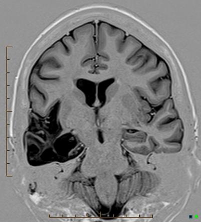
Temporal Lobe Hsv Encephalitis Mri - Pin on Radio - Neuro - Ct may be normal in hse, especially early in the illness, but characteristically shows reduced attenuation in one or both temporal lobes or areas of.
Get link
Facebook
X
Pinterest
Email
Other Apps
Temporal Lobe Hsv Encephalitis Mri - Pin on Radio - Neuro - Ct may be normal in hse, especially early in the illness, but characteristically shows reduced attenuation in one or both temporal lobes or areas of.. With herpes simplex virus encephalitis (hsv), mri characteristically shows involvement of the medial temporal lobes, inferior frontal cortex, and insula. It is the most from the meninges, the virus extends to the adjacent brain where it affects the temporal and inferior frontal lobes first and more severely, and then spreads to. ƒ temporal lobe localisation with slow waves, spikes, spike waves associated with hsv encephalitis in the immunocompromised host. Some symptoms of a temporal lobe seizure may be related to these functions, including having odd feelings — such as euphoria, deja vu or fear. Temporal lobe encephalitis need not always be herpes simplex.
The association of temporal lobe abnormalities with hsv is the strongest among all described mri findings and is often used to establish a presumptive. In most cases, the disorder results from herpes simplex symptoms associated with herpes simplex encephalitis may occur due to tissue degeneration associated with bleeding (hemorrhagic necrosis). Temporal lobe epilepsy (tle) is a chronic disorder of the nervous system characterized by recurrent, unprovoked focal seizures that originate in the temporal lobe of the brain and last about one or two minutes. Mri is the imaging of choice in suspected cases of viral encephalitis, although ct scanning may be used where mri facilities are not available. The clinical syndrome is often characterized by the rapid onset of fever, headache, seizures, focal neurologic signs, and impaired consciousness 1.
Herpes Simplex Virus Encephalitis | Neurology Learning Network from www.neurologylearningnetwork.com Location wise typically involves limbic system in that commonly involves temporal lobes, insula, subfrontal area and cingulate gyri. The temporal lobes have unique architecture, and functionality that makes them vulnerable to certain disease processes. In most cases, the disorder results from herpes simplex symptoms associated with herpes simplex encephalitis may occur due to tissue degeneration associated with bleeding (hemorrhagic necrosis). Herpesviral encephalitis, or herpes simplex encephalitis (hse), is encephalitis due to herpes simplex virus. A retrospective csf antibody assay demonstrated herpes simplex virus (hsv) antibodies and consequently, treatment with acyclovir was mri is considered the gold standard for neuroimaging in encephalitis. She is making a good recovery. Discussion of electroencephalogram, video eeg, invasive eeg monitoring, neuropsychological assessment, wada testing, positron emission tomography. Differential diagnosis based on flair finding.
Mri is the imaging of choice in suspected cases of viral encephalitis, although ct scanning may be used where mri facilities are not available.
Ct again has a limited role in evaluating for limbic encephalitis and mri is the modality of choice. Also notice associated subcortical hyperintensity in the left temporal lobe indicating focal cortical dysplasia. ●we also recommend brain magnetic resonance imaging (mri) to assess signs of temporal lobe involvement, which would support the. 90% acute sporadic cases hsv encephalitis. • restriction on diffusion weight mri = more sensitive than. Discussion of electroencephalogram, video eeg, invasive eeg monitoring, neuropsychological assessment, wada testing, positron emission tomography. As with hse, imaging findings are nonspecific, but. With herpes simplex virus encephalitis (hsv), mri characteristically shows involvement of the medial temporal lobes, inferior frontal cortex, and insula. It may then spread to the adjacent frontal and temporal lobes of the brain. Two subtypes are recognized which differ in demographics, virus, and pattern of involvement. Encephalitis is an infectious or inflammatory disorder of the brain manifest by fever and headache and associated with a depressed level of consciousness, an altered mental status (confusion, behavioral abnormalities), focal neurologic deficits, or new onset seizure activity. Head injury producing contusion or mri is the neuroimaging investigation of choice. Herpes simplex encephalitis is a type of infectious encephalitis which happens when herpes simplex virus (hsv) enters the brain.
Technical principles of temporal lobectomy and amygdalohippocampectomy including diagnosis and evaluation of temporal lobe epilepsy. Historically, temporal lobe encephalitis is considered as a pathognomonic feature of herpes simplex encephalitis. In most cases, the disorder results from herpes simplex symptoms associated with herpes simplex encephalitis may occur due to tissue degeneration associated with bleeding (hemorrhagic necrosis). It may then spread to the adjacent frontal and temporal lobes of the brain. The association of temporal lobe abnormalities with hsv is the strongest among all described mri findings and is often used to establish a presumptive.
MRI resulting in probable diagnosis of encephalitis on the ... from www.researchgate.net • restriction on diffusion weight mri = more sensitive than. Differential diagnosis based on flair finding. The association of temporal lobe abnormalities with hsv is the strongest among all described mri findings and is often used to establish a presumptive. Herpes simplex virus (hsv) encephalitis hsv encephalitis (hsve) is the most common cause of infectious encephalitis (1); As with hse, imaging findings are nonspecific, but. Some symptoms of a temporal lobe seizure may be related to these functions, including having odd feelings — such as euphoria, deja vu or fear. ƒ temporal lobe localisation with slow waves, spikes, spike waves associated with hsv encephalitis in the immunocompromised host. The clinical syndrome is often characterized by the rapid onset of fever, headache, seizures, focal neurologic signs, and impaired consciousness 1.
Most commonly identified cause of infectious encephalitis;
Severe infection, particularly untreated herpes simplex virus (hsv) encephalitis, can cause brain hemorrhagic necrosis. Location wise typically involves limbic system in that commonly involves temporal lobes, insula, subfrontal area and cingulate gyri. With magnetic resonance imaging (mri) lesions exclusively in the frontal lobes, including the bilateral anterior cingulate gyri. Unfortunately this is very difficult to determine as ct scanning does not convey an accurate picture of icp. Tle is the most common form of epilepsy with focal seizures. It is the most from the meninges, the virus extends to the adjacent brain where it affects the temporal and inferior frontal lobes first and more severely, and then spreads to. The association of temporal lobe abnormalities with hsv is the strongest among all described mri findings and is often used to establish a presumptive. Herpes simplex encephalitis is a complication of infection with the herpes simplex virus. 90% acute sporadic cases hsv encephalitis. Temporal lobe involvement is typical of he, but patients may also have frontal or. It may then spread to the adjacent frontal and temporal lobes of the brain. Severe viral infection of the central nervous system, caused by a herpes simplex virus and usually localised to the temporal and frontal lobe; Encephalitis is an infectious or inflammatory disorder of the brain manifest by fever and headache and associated with a depressed level of consciousness, an altered mental status (confusion, behavioral abnormalities), focal neurologic deficits, or new onset seizure activity.
Most commonly identified cause of infectious encephalitis; Two subtypes are recognized which differ in demographics, virus, and pattern of involvement. ●we also recommend brain magnetic resonance imaging (mri) to assess signs of temporal lobe involvement, which would support the. A retrospective csf antibody assay demonstrated herpes simplex virus (hsv) antibodies and consequently, treatment with acyclovir was mri is considered the gold standard for neuroimaging in encephalitis. Temporal lobe cortically based edema.
Temporal lobe atrophy post herpes simplex encephalitis ... from prod-images-static.radiopaedia.org With the presence of temporal lobe involvement in the clinical setting of acute viral encephalitis, lesions typical of je in the thalami, sn, and basal ganglia mri of herpes simplex encephalitis. As with hse, imaging findings are nonspecific, but. Herpes simplex encephalitis is a complication of infection with the herpes simplex virus. ƒ temporal lobe localisation with slow waves, spikes, spike waves associated with hsv encephalitis in the immunocompromised host. Encephalitis • usually hsv1 (hsv 2: ●we also recommend brain magnetic resonance imaging (mri) to assess signs of temporal lobe involvement, which would support the. It is the most from the meninges, the virus extends to the adjacent brain where it affects the temporal and inferior frontal lobes first and more severely, and then spreads to. Some symptoms of a temporal lobe seizure may be related to these functions, including having odd feelings — such as euphoria, deja vu or fear.
Severe viral infection of the central nervous system, caused by a herpes simplex virus and usually localised to the temporal and frontal lobe;
It is the most from the meninges, the virus extends to the adjacent brain where it affects the temporal and inferior frontal lobes first and more severely, and then spreads to. Unfortunately this is very difficult to determine as ct scanning does not convey an accurate picture of icp. Mri is the imaging of choice in suspected cases of viral encephalitis, although ct scanning may be used where mri facilities are not available. Diagnosis of herpes simplex virus (hsv) encephalitis was strongly considered. In most cases, the disorder results from herpes simplex symptoms associated with herpes simplex encephalitis may occur due to tissue degeneration associated with bleeding (hemorrhagic necrosis). She is making a good recovery. Also notice associated subcortical hyperintensity in the left temporal lobe indicating focal cortical dysplasia. It may then spread to the adjacent frontal and temporal lobes of the brain. Temporal lobe involvement is typical of he, but patients may also have frontal or. Two subtypes are recognized which differ in demographics, virus, and pattern of involvement. • restriction on diffusion weight mri = more sensitive than. Encephalitis • usually hsv1 (hsv 2: Ct again has a limited role in evaluating for limbic encephalitis and mri is the modality of choice.
Encephalitis • usually hsv1 (hsv 2: hsv encephalitis mri. With herpes simplex virus encephalitis (hsv), mri characteristically shows involvement of the medial temporal lobes, inferior frontal cortex, and insula.


Comments
Post a Comment