Nasal Cavity Bones Anterior View - Nasal Bone Wikipedia - — drains into nasal cavity gives us mucus 2.
Get link
Facebook
X
Pinterest
Email
Other Apps
Nasal Cavity Bones Anterior View - Nasal Bone Wikipedia - — drains into nasal cavity gives us mucus 2.. Inside the nasal area of the skull, the nasal cavity is divided into halves by the nasal septum. Anterior view of the skull. The bones associated with the skull including the ear ossicles and hyoid bones. For more anatomy content please follow us and visit our website: The cribriform foramina of the ethmoid bone.
The exterior nares, or nostrils lead into this portion of the nasal cavity which is essentially just a short passageway lined with hair that leads into the respiratory region of the nasal cavity. This makes the skull very strong. The cribriform foramina of the ethmoid bone. Which landmark is not visible from an anterior view? Which bone forms the anterior cranium?
7 2 The Skull Medicine Libretexts from med.libretexts.org Superior wall of nasal cavities. Anatomynote.com found oral cavity anterior view and lateral view from plenty of anatomical pictures on the internet. They lie side by side and are fused at the midline to form the bridge of the nose (nasal septum). What part of the nasal cavity is formed by the nasal septum? The cribriform foramina of the ethmoid bone. 4 the nasal vestibules are the two entry points into the nasal cavity. The nasal cavity is divided into three regions: The nasal cavity is formed by the vomer and the nasal, lachrymal, and turbinate bones.
Identify the part of the ethmoid bone that contributes to the nasal septum.
The bones associated with the skull including the ear ossicles and hyoid bones. Lateral, major alar, minor alar, and the cartilaginous septum.the lateral and major alar cartilages are the largest, and contribute the most to the. The structure is also referred to as the piriform aperture. In the anterior nasal cavity there are nasoturbinates and maxilloturbinates (figure 2.3.60), whereas the posterior nasal cavity contains ethmoturbinates.among the turbinates are the air passages, the dorsal, middle and. These are formed by bone and covered by mucous membrane. The two nasal cavities sit within the external nose and the adjacent skull. Nasal part of frontal bone; The cribriform foramina of the ethmoid bone. The inferior portion of the nose is made up of hyaline cartilages; This makes the skull very strong. Inside the nasal area of the skull, the nasal cavity is divided into halves by the nasal septum. The exterior nares, or nostrils lead into this portion of the nasal cavity which is essentially just a short passageway lined with hair that leads into the respiratory region of the nasal cavity. The walls of the nasal cavity are formed by several bones of the skull, and the posterior and anterior walls open with large apertures.
Which bone forms the anterior cranium? It extends from the vestibule of the nose to the nasopharynx, and has three divisions: The nasal cavity is divided into two lateral compartments separated down the middle by the nasal septum. Inside the nasal area of the skull, the nasal cavity is divided into halves by the nasal septum. The frontal bone articulates with the parietal bone by means of the sagittal suture.
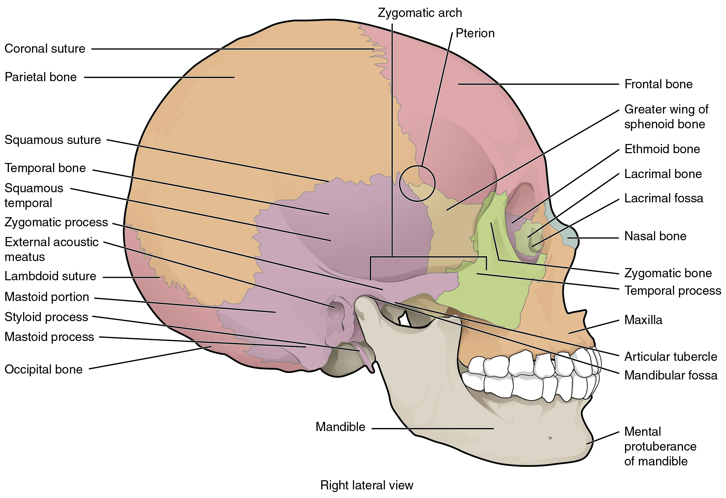
Human Being Anatomy Skeleton Lateral View Of Skull Image Visual Dictionary Online from www.visualdictionaryonline.com These are formed by bone and covered by mucous membrane. The nasal bones are small oblong bones somewhat rectangular in shape. Maxillary sinuses, frontal sinuses, sphenoidal sinuses, and ethmoidal. Posteriorly the cavities communicate with the nasopharynx by two apertures called choanae. The walls of the nasal cavity are formed by several bones of the skull, and the posterior and anterior walls open with large apertures. Inside the nasal area of the skull, the nasal cavity is divided into halves by the nasal septum. Lateral view of skull a view of the lateral skull is dominated by the large, rounded brain case above and the upper and lower jaws with their teeth below (figure 6.18). Mandible bone marking in anterior view of skull 1.
Anterior view of the skull.
I.levels of organisation, homeostasis and nomenclature The nasal vestibule, the nasal cavity proper or nasal fossa, and the olfactory region. The nasal cavity is the most superior part of the respiratory tract. Figure 7.13 lateral wall of nasal cavity the three nasal conchae are curved bones that project from the lateral walls of the nasal cavity. Other animals edit in primitive bony fish and tetrapods , the nasal bones are the most anterior of a set of four paired bones forming the roof of the skull , being followed in sequence by the frontals. Three cartilages contribute to the nasal septum: Paranasal sinuses have four different pairs: For more anatomy content please follow us and visit our website: 4 the nasal vestibules are the two entry points into the nasal cavity. The anterior nasal aperture is simply the area where the anterior bony aspects of both the maxilla and the nasal bone terminate and form an opening into the cartilaginous nasal vestibule. The nasal bones are small oblong bones somewhat rectangular in shape. Anterior and external view of nasal cavity. Two of the cranium, the frontal and ethmoid, and two of the face, the opposite nasal and the maxilla.
An anterior view of the skull shows the bones that form the forehead, orbits (eye sockets), nasal cavity, nasal septum, and upper and lower jaws. Mandible bone marking in anterior view of skull 1. The nasal cavity is separated by a cartilaginous septum. Endoscopic view through the anterior nasal aperture. The nasal cavity is divided into three regions:
Supportive Bones And Cartilages Of The Nasal Cavity from www.getbodysmart.com The nasal articulates with four bones: This makes the skull very strong. The anterior nasal aperture is simply the area where the anterior bony aspects of both the maxilla and the nasal bone terminate and form an opening into the cartilaginous nasal vestibule. An anterior view of the skull shows the bones that form the forehead, orbits (eye sockets), nasal cavity, nasal septum, and upper and lower jaws. In infants the sutures (joints) between the various skull elements are loose, but with age they fuse together. How many bones make up. Anatomynote.com found oral cavity anterior view and lateral view from plenty of anatomical pictures on the internet. Three cartilages contribute to the nasal septum:
Two of the cranium, the frontal and ethmoid, and two of the face, the opposite nasal and the maxilla.
Identify the part of the ethmoid bone that contributes to the nasal septum. An anterior view of the skull shows the bones that form the forehead, orbits (eye sockets), nasal cavity, nasal septum, and upper and lower jaws. The anterior nasal aperture is simply the area where the anterior bony aspects of both the maxilla and the nasal bone terminate and form an opening into the cartilaginous nasal vestibule. The nasal cavity communicates anteriorly through the nostrils and posteriorly with the nasopharynx through openings called choanae. What part of the nasal cavity is formed by the nasal septum? The lower parts of the nasal bones, the cartilage attached to them, the outer nose, and the nostrils are there in the front part of the nasal cavity. Paranasal sinuses have four different pairs: The bones of the skull slot together like a jigsaw puzzle. Lateral view of skull a view of the lateral skull is dominated by the large, rounded brain case above and the upper and lower jaws with their teeth below (figure 6.18). In infants the sutures (joints) between the various skull elements are loose, but with age they fuse together. Maxillary sinuses, frontal sinuses, sphenoidal sinuses, and ethmoidal. Each is a triangular space situated anterior to the limen nasi and defined laterally by the alae nasi, medially by the membranous septum. The nasal cavity receives innervation from both external and internal carotid arteries.
G, anterior endoscopic view of reconstructed sphenoid, ethmoid, and both palatine bones nasal cavity bones. The nasal cavity is the most superior part of the respiratory tract.


Comments
Post a Comment